
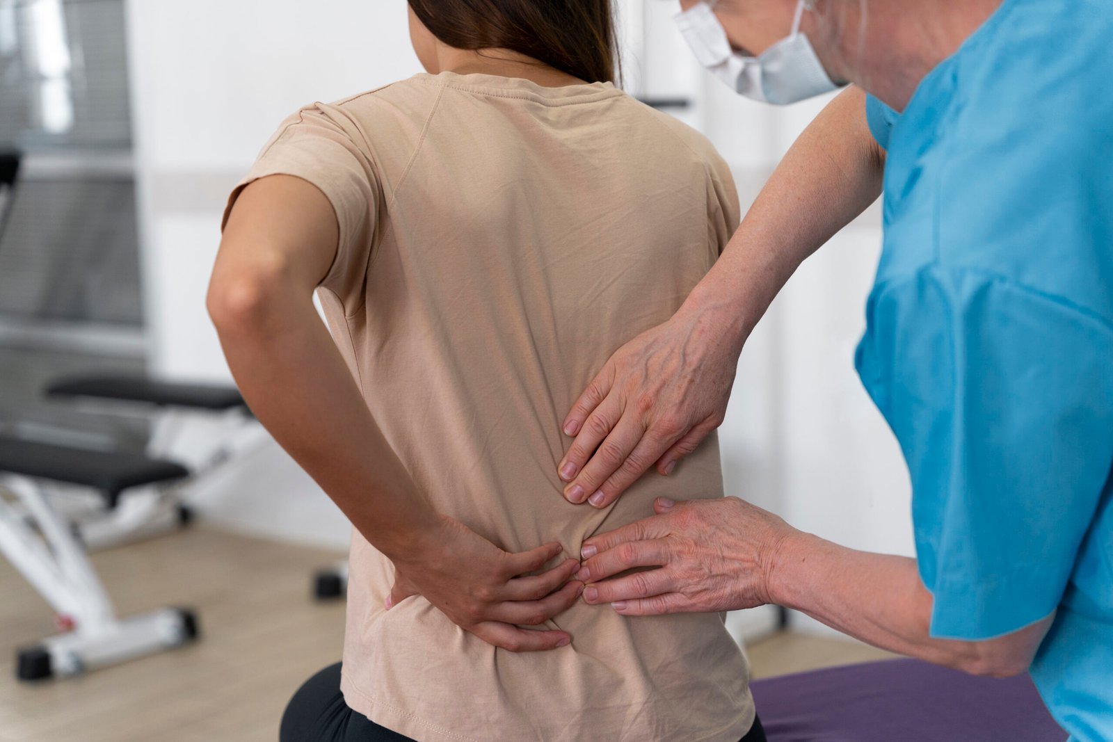
Spinal Stenosis: Causes, symptoms and treatment options
Spinal stenosis is a common spinal disorder that affects millions of adults worldwide, especially as the human spine undergoes natural changes over time. When narrowing occurs within the spaces of the backbone, it places unwanted mechanical pressure on the spinal cord and the nerves traveling through the canal. This narrowing can lead to discomfort, restricted mobility, and persistent nerve pain that disrupts everyday routines. Understanding how this condition develops, recognizing its early warning signs, and reviewing modern medical pathways empowers individuals to take charge of their spinal health and find long-term relief.
What is Spinal Stenosis and how does it develop?
The spine consists of a stacked column of bones called vertebrae, separated by shock-absorbing intervertebral discs. Running directly down the center of this bony column is the spinal canal, a protected passageway housing the spinal cord and a complex network of branching nerve roots.
Spinal stenosis occurs when one or more areas within this canal begin to narrow. As the available space diminishes, the physical structure pinches or compresses the neural tissue inside. Medical professionals classify spinal stenosis primarily by where the narrowing takes place along the spinal column:
- Lumbar spinal stenosis: This is the most widespread variety. It occurs in the lower back and frequently compresses the nerve roots that extend down into the hips, buttocks, thighs, and calves.
- Cervical spinal stenosis: This form develops in the neck. Because the neck canal encloses the upper spinal cord itself, compression in this area can cause symptoms extending into the shoulders, arms, hands, and occasionally the legs.
- Thoracic spinal stenosis: Narrowing in the mid-back region is far less common, though it can still happen following trauma or specific structural shifts.
Because spinal stenosis usually advances at an incremental pace, the earliest structural changes often go unnoticed for years before creating functional difficulties.
Common causes and risk factors behind spinal stenosis
Most cases of spinal stenosis are acquired rather than present at birth, developing as a natural consequence of the aging process and structural wear and tear. Over decades, daily biomechanical stress alters the joints, ligaments, and bones of the vertebral column.
The primary underlying causes include:
- Osteoarthritis and Bone Spurs: As the cartilage that cushions facet joints breaks down, bone rubs directly against bone. The body responds to this friction by producing extra bone growth known as osteophytes or bone spurs. When these smooth bony projections grow inward toward the spinal canal, they narrow the passage and trigger spinal stenosis.
- Herniated or Bulging Discs: With age, the spinal discs dry out, lose their height, and become brittle. When a disc tears or bulges outward into the canal, it directly reduces the space reserved for the nervous system.
- Thickened Ligaments: The tough fibrous bands that connect and stabilize adjacent vertebrae, such as the ligamentum flavum, can stiffen and thicken over time. This tissue hypertrophy bulges into the canal, contributing substantially to spinal stenosis.
- Spondylolisthesis: This condition occurs when one vertebra slips forward over the one beneath it, throwing the canal out of alignment and squeezing the nerve passage.
- Spinal Trauma and Fractures: Accidents, sports collisions, or falls can crack or dislocate vertebrae, producing sharp bone fragments or tissue inflammation that crowd the canal.
- Congenital Narrowing: A small percentage of individuals are born with a naturally narrow canal, meaning mild wear and tear later in life can produce symptomatic spinal stenosis at an earlier age.
Recognizing the Symptoms: From dull aches to radiating pain
Because the narrowing process happens gradually, spinal stenosis symptoms usually appear progressively rather than all at once. The specific discomfort depends entirely on whether the compression is located in the neck or the lower back.
Lumbar Symptoms
When spinal stenosis strikes the lumbar region, the defining symptom is neurogenic claudication. This term describes cramping, heaviness, or numbness in the buttocks and legs that flares up during standing or walking. Individuals frequently notice that sitting down, resting, or leaning forward over a shopping cart relieves the pain almost immediately. Leaning forward flexes the lumbar spine, which temporarily opens the canal and relieves pressure on compressed nerves. Other common lower back signs include:
- Burning or electric shock sensations radiating down the back of the leg (sciatica)
- Pins-and-needles sensations in the feet or toes
- Muscle fatigue and weakness in the legs after walking short distances
- A feeling of instability when upright
Cervical Symptoms
When cervical spinal stenosis compresses the spinal cord (a condition known as cervical myelopathy), the symptoms can be subtle at first but carry significant consequences if neglected. Typical signs include:
- Numbness, tingling, or weakness in the hands, fingers, or forearms
- Difficulty with fine motor tasks, such as buttoning a shirt, tying shoelaces, or holding a pen
- Frequent dropping of everyday objects
- Balance disturbances, unsteadiness, or a clumsy, staggering gait
- Neck stiffness and radiating shoulder discomfort
How physicians diagnose spinal stenosis
An accurate diagnosis begins with a thorough clinical evaluation. A spine specialist reviews medical history, asks detailed questions regarding when and where pain strikes, and conducts physical and neurological assessments. During the exam, the doctor evaluates muscle strength, sensory perception, reflexes, walking balance, and how posture changes symptom intensity.
To confirm the presence and severity of spinal stenosis, medical teams rely on modern diagnostic imaging:
- Magnetic Resonance Imaging (MRI): The gold standard for evaluating soft tissues, an MRI provides detailed cross-sectional views of the spinal cord, exiting nerve roots, intervertebral discs, and thickened ligaments.
- Computed Tomography (CT) Scan or CT Myelogram: When an MRI cannot be performed, a CT scan offers high-resolution bone images. Pairing it with a contrast dye injection (myelogram) outlines the spinal canal to reveal nerve compression clearly.
- Digital X-rays: While standard X-rays cannot visualize nerves or soft discs, they highlight bone spurs, disc height loss, overall alignment, and spinal instability.
Non-surgical treatment options for daily relief
The vast majority of people diagnosed with spinal stenosis manage their symptoms successfully through conservative, non-operative therapies. The goal of non-surgical care is to reduce inflammation, alleviate nerve compression, and restore functional mobility.
Conservative management typically combines several modalities:
- Targeted Physical Therapy: A specialized physical therapist teaches exercises that strengthen abdominal and back core muscles, improve flexibility, and encourage flexion postures. Strengthening the supportive core structures unloads excess pressure from the spine.
- Activity Modification: Adjusting everyday habits provides fast symptomatic comfort. Switching from walking to stationary cycling, taking frequent sitting breaks, and maintaining a healthy body weight reduce direct stress on the spine.
- Medications: Over-the-counter pain relievers, such as nonsteroidal anti-inflammatory drugs, help reduce surrounding tissue swelling. For nerve-related pain, physicians may prescribe neuropathic agents or short courses of muscle relaxants to ease associated spasms.
- Epidural Steroid Injections: When oral medications and physical therapy do not offer sufficient relief, an image-guided epidural injection can deliver a concentrated corticosteroid directly into the space surrounding the compressed nerves. This treatment helps reduce local swelling and calms nerve irritation for weeks or months at a time.
Might interest you: Can scoliosis get worse in adulthood?
Surgical solutions and advanced spine care
When conservative therapies no longer provide adequate pain relief, or if severe nerve compression causes progressive muscle weakness, numbness, or loss of balance, surgical intervention becomes an important consideration. Modern spine surgery focuses on decompressing the pinched neural elements while preserving as much healthy bone and tissue as possible.
Common surgical procedures include:
- Decompressive Laminectomy: The surgeon removes the lamina (the bony roof of the vertebra) and any overgrown bone spurs or thickened ligaments to expand the canal diameter and free the trapped nerves.
- Foraminotomy: This technique widens the small neural exit holes (foramina) where nerve roots branch away from the main spinal canal.
- Spinal Fusion: If spinal stenosis is accompanied by spondylolisthesis or significant structural instability, fusing two or more vertebrae together with bone grafts and titanium hardware stabilizes the spine and prevents painful abnormal motion.
- Minimally Invasive Spine Surgery: Whenever appropriate, surgeons utilize specialized tubular retractors, tiny incisions, and high-definition microscopes or endoscopes. This approach minimizes muscle disruption, reduces postoperative discomfort, and shortens hospital recovery times.
For patients seeking precise diagnosis, tailored non-operative plans, or advanced surgical solutions for spinal stenosis, consulting an experienced, specialized spine surgeon is essential. Leading Spine Surgeon, Dr. Konstantinos Starantzis, provides expert clinical evaluation and state-of-the-art care, applying the latest evidence-based techniques to help patients regain mobility, overcome persistent nerve pain, and return to an active, fulfilling life.
Stay informed with Athens Pulse.